Visual Evoked Potentials (VEP) is a specialized neurophysiological test used to measure the electrical activity of the brain in response to visual stimulation. The test evaluates how effectively visual signals travel from the eyes through the optic nerves to the visual centers of the brain. When this pathway is affected by inflammation, compression, injury, or neurological disease, the transmission of signals may be delayed or reduced. Visual Evoked Potentials help detect such abnormalities with precision, even when imaging studies appear normal.
Dr. Raghu Samala utilizes VEP as part of a comprehensive neurological evaluation to assess visual pathway integrity. The test is non-invasive, safe, and provides valuable diagnostic information in patients experiencing unexplained vision problems or suspected neurological disorders affecting the optic nerve.
Abnormal Visual Evoked Potentials may be associated with conditions that disrupt the optic nerve or visual pathways within the brain. Common causes include optic neuritis, multiple sclerosis, brain tumors affecting the visual pathways, head injuries, stroke, and compressive lesions. In some cases, metabolic or inflammatory disorders can also interfere with signal transmission.
Early detection of abnormalities in the visual pathway is crucial, especially in progressive neurological conditions, as timely intervention can help prevent further deterioration.
Patients who are advised to undergo a VEP study often report blurred vision, double vision, reduced visual clarity, partial loss of vision, or changes in color perception. Some individuals may experience pain with eye movement or sudden visual disturbances without an obvious cause.
In certain neurological disorders, visual symptoms may be the first indication of an underlying condition. When visual changes persist or are accompanied by other neurological symptoms such as weakness, numbness, or imbalance, further evaluation becomes essential.
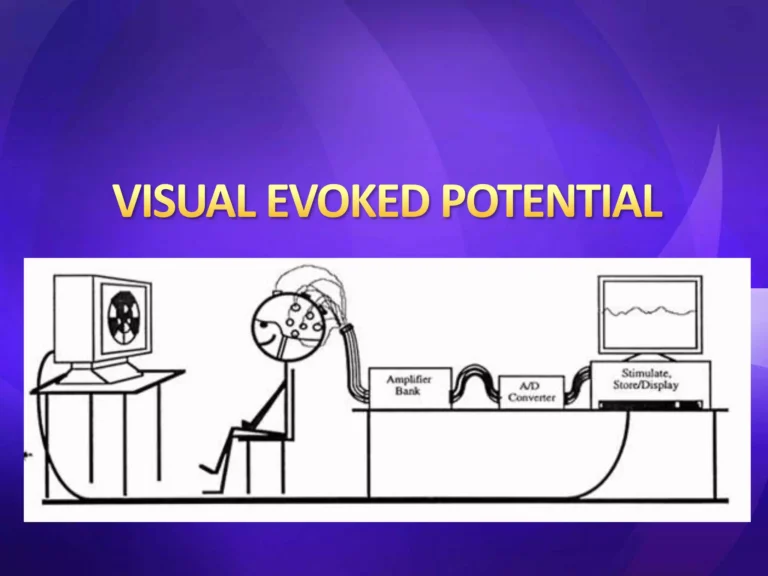
During a Visual Evoked Potentials test, small electrodes are placed on the scalp over the visual cortex at the back of the head. The patient is asked to look at a visual stimulus, usually a patterned screen or flashing light. The brain’s electrical response to this visual input is recorded and analyzed. The test measures the speed and strength of signal transmission along the visual pathways.
The procedure is painless and typically completed within a short duration. Dr. Raghu Samala carefully evaluates the recorded responses to identify delays or abnormalities that may indicate optic nerve or central nervous system dysfunction. When combined with clinical examination and imaging studies such as MRI, VEP provides a comprehensive assessment of visual pathway health.
Treatment depends on the underlying cause identified through the evaluation. Inflammatory conditions such as optic neuritis may be managed with appropriate medications to reduce inflammation. If a compressive lesion such as a tumor is detected, surgical intervention may be considered. In cases of multiple sclerosis or other demyelinating disorders, targeted medical therapy is initiated to manage disease progression.
Dr. Raghu Samala develops individualized treatment plans aimed at preserving vision, addressing the root cause, and improving overall neurological function.
After treatment, regular follow-up is important to monitor recovery and ensure stabilization of visual function. Patients may be advised to attend periodic neurological evaluations and repeat diagnostic testing if required. Managing underlying systemic conditions and adhering to prescribed medications play a key role in long-term outcomes.
Rehabilitation strategies, including vision therapy in selected cases, may also support recovery and adaptation.
Visual Evoked Potentials is a safe and non-invasive diagnostic procedure with minimal risks. The electrodes simply record brain activity and do not deliver electrical currents to the brain. Some patients may experience mild eye strain during visual stimulation, but this is temporary.
The primary concern lies in delaying diagnosis, as untreated conditions affecting the visual pathways may lead to progressive vision loss.
You should seek medical evaluation if you experience unexplained vision changes, persistent blurred or double vision, sudden vision loss, or visual symptoms accompanied by other neurological signs. Early assessment is especially important if symptoms develop suddenly or worsen over time.
Timely diagnosis through Visual Evoked Potentials allows accurate identification of visual pathway disorders and helps guide effective treatment. If you are experiencing concerning visual or neurological symptoms, consultation with Dr. Raghu Samala ensures expert evaluation and comprehensive care tailored to your needs.